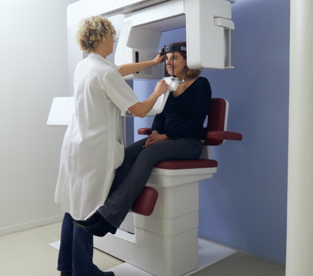
In our practice, we use a three-dimensional radiodiagnostic device (Cone Beam) to diagnose disturbance fields and to safely plan the placement of implants.
This radiological examination allows us to see the bone in great detail.En particulier, il nous est possible de visualiser au bout de racines de dents dévitalisées des infections et des kystes qui ne sont pas visibles sur les clichés panoramiques ou rétro alvéolaire.Il nous permet de visualiser les zones d’ostéolyse, de voir les corps étrangers et de repérer toutes les structures anatomiques qu’il est nécessaire de protéger pendant notre chirurgie.It is necessary to perform 3D implant imaging to identify the bone volume available, especially when inserting implants directly after the extraction of a tooth.NICOs are also identified by visualising the absence of bone density on 3D imaging. We can then measure the height, the length, the width, of the NICO and understand its relation to the sensitive anatomical zones like the inferior alveolar nerve or the upper maxillary sinus cavity which makes it possible to protect them more effectively.